

Excess parathyroid hormone drives high-turnover bone loss that hits cortical bone hardest, raising fracture risk even when spine scans look normal. Curative parathyroidectomy usually reverses much of that damage, with measurable bone density gains starting around 6 months and continuing for up to 2 years. When surgery isn’t an option, medications and vitamin D management can still protect or improve bone density.
TL;DR:
- Cortical bone loss in hyperparathyroidism occurs mainly at the wrist and hip, often without significant changes in lumbar spine scans.
- Standard DXA tests at the spine may underestimate fracture risk because they overlook cortical damage detectable through radius measurements or advanced imaging.
- Parathyroidectomy typically results in measurable bone density gains at the spine and hip starting around six months post-surgery, with about 40% of patients experiencing over 5% improvement.
- Medical treatments like bisphosphonates and denosumab can slow bone loss but do not fully reverse cortical damage or replace surgical intervention.
- Early and comprehensive monitoring, including three-site DXA, TBS, or HR-pQCT, enhances fracture risk assessment and guides optimal timing for treatment decisions.
Table of Contents
- How Hyperparathyroidism Affects Bone Density and Fracture Risk
- How Doctors Actually Measure Bone Health When PTH Is High
- Does Parathyroidectomy Improve Bone Density? What the Data Actually Shows
- Non-Surgical Treatments: What Medications Can and Can’t Do for Bone
- Who Gains the Most Bone Density After Surgery?
- Monitoring, Supplements, and Exercise: A Realistic Plan Before and After Treatment
- Where Supervised Bone-Loading Sessions Fit Alongside Medical Care
- Secondary Hyperparathyroidism: A Different Cause, A Different Fix
- What’s Actually Happening at the Cellular Level
- Fracture Risk Tools: Why Standard Calculators Can Miss the Danger
- Ruling Out Look-Alike Conditions
- What Happens If Hyperparathyroidism Goes Untreated
- A Checklist Worth Bringing to Your Next Appointment
- Supporting Your Bones Between Medical Visits
- Sources
- FAQ
How Hyperparathyroidism Affects Bone Density and Fracture Risk
Parathyroid hormone (PTH) is supposed to work in short bursts, nudging calcium out of bone only when the body needs it. Primary hyperparathyroidism breaks that rhythm. A parathyroid tumor, almost always benign, pumps out PTH continuously instead of in pulses, and that constant signal turns bone remodeling into a losing battle.
Here’s the mechanism in plain terms: continuous PTH exposure activates osteoclasts, the cells that break down bone, faster than osteoblasts can rebuild it. Bone turnover accelerates across the entire skeleton, but the resorption side wins the race. The result is a specific, recognizable pattern of damage rather than uniform thinning everywhere.
Cortical bone, the dense outer shell that makes up most of the radius, hip, and long bones, takes the brunt of it. It thins out and develops microscopic porosity, essentially becoming Swiss cheese at a level invisible to the naked eye. Trabecular bone, the spongy inner lattice found mostly in the vertebrae, sometimes appears deceptively stable on a standard scan.
That mismatch matters enormously for how hyperparathyroidism gets diagnosed and monitored. A patient can have a completely normal or even reassuring lumbar spine DXA score while cortical bone at the wrist or hip is quietly deteriorating. This is one of the more counterintuitive things about the condition: the site where doctors most often look first (the spine) is frequently the site least affected.
What this looks like in practice:
- Forearm and hip bone density typically drop first and drop hardest.
- Lumbar spine density can stay in the normal or osteopenic range for years, masking the real extent of loss.
- Fracture risk rises at wrist, hip, and vertebral sites, often before any single DXA score crosses into the osteoporosis threshold.
- Silent vertebral fractures can occur even in patients whose spine T-scores never trigger a diagnosis of osteoporosis on paper.
The disconnect between DXA scores and actual fracture risk is one of the most underappreciated features of primary hyperparathyroidism. Population studies have repeatedly found that people with PHPT fracture more often than their T-scores would predict, largely because standard central DXA undersells cortical bone loss. This is why an endocrinologist might order additional imaging even when a patient’s bone density numbers look unremarkable.
If you’ve been told your bone density is “fine” based on a spine scan alone, that finding deserves a second look once hyperparathyroidism enters the picture. The condition doesn’t behave like typical age-related osteoporosis, and it shouldn’t be assessed the same way.
How Doctors Actually Measure Bone Health When PTH Is High
Standard bone density testing wasn’t built with hyperparathyroidism’s cortical bias in mind, which is why clinicians increasingly reach for a broader toolkit.
- Dual-energy X-ray absorptiometry (DXA) at three sites. A complete workup includes the lumbar spine, total hip, and the 1/3 distal radius, not just the spine or hip alone. The radius site catches the cortical bone loss that spine-only scans routinely miss, and guidelines for primary hyperparathyroidism specifically call for including it.
- Trabecular Bone Score (TBS). TBS is calculated from the same spine DXA image but analyzes the texture of the bone rather than just its density. It can flag degraded microarchitecture in patients whose spine BMD looks normal, and it improves fracture-risk prediction specifically in cases where DXA results seem discordant with clinical risk.
- High-resolution peripheral quantitative computed tomography (HR-pQCT). This imaging technique, available mostly at academic or research centers, produces a three-dimensional picture of cortical and trabecular microarchitecture at the wrist and ankle. It’s not routine for every patient, but it’s the gold standard when researchers or specialists need to see bone quality, not just bone quantity.
- Bone turnover markers. Blood or urine markers like CTX (a resorption marker) and P1NP (a formation marker) reflect how fast bone is being broken down and rebuilt. They’re not diagnostic on their own, but tracking them before and after treatment can show whether therapy is working faster than a repeat DXA would reveal.
- Vertebral imaging. Because vertebral fractures in hyperparathyroidism can be silent, meaning they cause no acute pain, some clinicians order a vertebral fracture assessment or spine X-ray when clinical suspicion is high, even without a clear T-score trigger.
Pro Tip: If your DXA report only lists spine and hip numbers, ask whether a 1/3 distal radius measurement was included. It’s a small addition to the same appointment, and in hyperparathyroidism, it’s often the number that tells the real story.
None of these tools replace clinical judgment, and not every patient needs the full panel. A newly diagnosed patient with mild, asymptomatic disease and normal DXA results may need nothing beyond routine monitoring. Someone with a high PTH level, low bone density at any site, or a prior fragility fracture is a much stronger candidate for TBS or radial imaging before deciding on surgery versus watchful waiting.
Does Parathyroidectomy Improve Bone Density? What the Data Actually Shows
Surgical removal of the overactive parathyroid gland is the only treatment that addresses the root cause rather than managing its downstream effects, and the bone density evidence behind it is some of the most consistent data in endocrine surgery.
Multiple cohort studies tracking patients before and after parathyroidectomy show a clear, reproducible pattern: bone mineral density starts recovering by around 6 months post-surgery, with the biggest and most consistent gains showing up at the lumbar spine and hip, and continued improvement tracked out to 24 months. The timeline holds up across different patient populations, which is part of why endocrine surgery societies treat curative surgery as the default recommendation for eligible patients rather than one option among several.
The magnitude of recovery varies by site, and that variation tells you something important about how bone heals.
- Lumbar spine tends to show the fastest and largest percentage gains, since trabecular bone remodels faster than cortical bone.
- Femoral neck and total hip improve more gradually but consistently, with meaningful gains typically visible by the 1-year mark.
- Distal radius, the site hit hardest by the disease in the first place, recovers more slowly and less completely. Cortical bone that has thinned and become porous over years doesn’t rebuild as efficiently as trabecular bone, even after the hormonal driver is removed.
A prospective study tracking bone density changes across different hyperparathyroidism phenotypes found that roughly 40% of patients achieved a clinically meaningful gain, defined as more than 5% improvement, at the spine after surgery, with substantial proportions also crossing that threshold at the femur. That’s not universal, but it’s a strong enough signal that a large share of patients see recovery worth measuring, not just a modest uptick that rounds to noise.
High-resolution imaging adds a layer that DXA alone can’t provide. Studies using HR-pQCT have documented improvements in volumetric bone density, cortical thickness, and estimated bone strength beginning around the 6-month mark and continuing through 24 months. This matters because DXA measures density in two dimensions and can understate real structural recovery. When researchers modeled estimated bone strength directly from these 3D scans, the improvements tracked closely with the kind of architectural repair that should, in theory, translate into lower fracture risk, even in cases where the DXA percentage change alone looked unremarkable.
Here’s the number worth remembering: about 40% of surgical patients hit a >5% spine BMD gain, and gains at the hip and spine tend to continue accumulating for a full 2 years, not just the first 6 months.
What does this mean for someone weighing surgery against watchful waiting? A few realistic expectations are worth setting early:
- Don’t expect a dramatic jump on the first post-op scan at 6 months. It’s real, but it’s usually the beginning of a longer curve, not the peak.
- The spine will likely show the most encouraging numbers first. The hip catches up over the following year. The wrist may lag behind both, sometimes for years.
- A DXA at 12 months that shows only modest improvement doesn’t mean surgery failed. Bone remodeling operates on a biological clock that outlasts most patients’ patience.
- Not every patient sees a 5%+ gain, and that’s not necessarily a bad outcome. Stabilization after years of ongoing loss is itself a meaningful clinical win.
None of this is a guarantee. Age, kidney function, prior fracture history, and how long the hyperparathyroidism went untreated before surgery all shape how much bone actually comes back. But across the available prospective data, the trend line points the same direction: remove the source of excess PTH, and bone density measured at multiple sites tends to improve, with the clearest gains showing up in the first 2 years after the procedure.
Non-Surgical Treatments: What Medications Can and Can’t Do for Bone
Surgery isn’t always the immediate answer, whether because a patient has mild disease, isn’t a surgical candidate, or is still weighing the decision. Several medical options can protect or improve bone density in the meantime, though each comes with real limitations worth understanding before assuming a pill can substitute for a cure.
- Bisphosphonates (alendronate, risedronate, and similar drugs) slow osteoclast activity and can measurably increase BMD at the lumbar spine and hip in patients with hyperparathyroidism who don’t undergo surgery. They work best on trabecular-rich bone. Cortical bone, already thinned and porous from years of PTH excess, responds more slowly and less completely, since bisphosphonates suppress further resorption rather than actively rebuilding lost architecture.
- Denosumab, a monoclonal antibody given by injection, can produce meaningful gains in both BMD and TBS in patients with hyperparathyroidism, and some clinicians consider it when surgery isn’t feasible or a patient has already failed bisphosphonate therapy. It doesn’t fully reverse pre-existing cortical damage either, and stopping the drug carries a well-documented rebound risk, meaning bone loss can accelerate rapidly if a follow-up dose is missed or discontinued without a transition plan.
- Cinacalcet works differently. It lowers calcium levels by making the parathyroid gland’s calcium sensors more sensitive, which reduces PTH secretion. It’s genuinely useful for controlling dangerous hypercalcemia, but it isn’t primarily a bone-building drug, and its effect on BMD is far less consistent than what’s seen with antiresorptive therapy or surgery.
- Vitamin D repletion sits in a category of its own. Correcting a deficiency is important for skeletal health, but it has to be coordinated with endocrine care in hyperparathyroidism specifically, because raising vitamin D levels can sometimes worsen hypercalcemia in a patient whose parathyroid gland is already overactive. This is not a supplement to manage on your own.
Pro Tip: If you’re on cinacalcet for calcium control, don’t assume your bone density is automatically protected. Ask your endocrinologist directly whether an antiresorptive should be added, since the two drugs address different parts of the problem.
None of these medical options replace the biological reset that comes from removing the source of excess hormone. They’re genuinely useful tools for patients who can’t have surgery, who are waiting for a procedure, or whose disease is mild enough that observation with medical support is the appropriate call. But framing them as equivalent to a cure oversells what the evidence actually supports.
Who Gains the Most Bone Density After Surgery?
Not every surgical patient sees the same result, and a few patterns show up consistently enough across the research to be worth discussing with your care team before deciding on timing.
- Higher baseline PTH levels predict bigger gains. Patients who enter surgery with more severely elevated PTH, and who see larger drops in urinary calcium after their parathyroid level normalizes, tend to show larger BMD improvements at both the spine and femur afterward. In a sense, the sicker the bone was going in, the more room it has to recover.
- Phenotype matters less than you’d expect. Hyperparathyroidism shows up in a few different biochemical patterns: classic hypercalcemic disease, normocalcemic PHPT (calcium stays normal while PTH runs high), and normohormonal disease (PTH looks normal but calcium runs high). Studies comparing these phenotypes have found that surgery-related BMD gains occur across all three, and the differences between them tend to be modest rather than dramatic. Phenotype alone shouldn’t discourage anyone from considering surgery.
- Chronic kidney disease complicates the picture. Reduced kidney function affects vitamin D activation and calcium handling independently of the parathyroid tumor itself, which can blunt or delay how much bone density recovers after surgery.
- Severe pre-existing vitamin D deficiency can slow recovery until it’s corrected, since vitamin D is required for the mineralization that rebuilding bone depends on.
- Age and prior antiresorptive therapy both shape the trajectory. Older patients and those who’ve already been on bisphosphonates or denosumab for years may see a different recovery curve than a younger, treatment-naive patient, sometimes smaller in percentage terms simply because there’s less untreated damage left to reverse.
None of these factors should be read as reasons to avoid surgery. They’re more useful as a way to calibrate expectations. A 70-year-old with longstanding CKD and severe vitamin D deficiency is a legitimate surgical candidate, but their 12-month DXA follow-up conversation will likely look different from a 45-year-old with none of those complications.
Monitoring, Supplements, and Exercise: A Realistic Plan Before and After Treatment
Whether you’re heading toward surgery, managing hyperparathyroidism medically, or still working through a diagnosis, a few practical steps make the difference between guessing and actually tracking progress.
- Get a complete baseline DXA before treatment starts. That means spine, hip, and the 1/3 distal radius, not just one or two sites. Without this baseline, there’s nothing solid to compare later scans against.
- Schedule the first follow-up DXA at 6 to 12 months post-treatment, whether that treatment is surgery or a medication regimen. Earlier than 6 months, bone remodeling usually hasn’t produced a change large enough to detect reliably.
- Ask about TBS or HR-pQCT if fracture risk still feels uncertain after standard DXA results come back, particularly if your spine score looks better than your clinical history suggests it should.
- Coordinate vitamin D supplementation with your endocrinologist rather than self-dosing. The right target range and monitoring frequency depend on your calcium levels and whether your parathyroid disease is primary or secondary.
- Avoid unsupervised high-dose calcium supplements. In a condition already defined by calcium dysregulation, adding extra calcium without medical guidance can do more harm than good.
- Discuss whether to continue, pause, or start antiresorptive therapy around the time of surgery. Some patients benefit from bridging medication while awaiting a procedure; others need a clear plan for what happens to that medication afterward.
- Build in supervised weight-bearing and resistance exercise. Mechanical loading is one of the few inputs that reliably stimulates bone formation, and it complements whatever medical or surgical treatment is already underway. Programs that use structured, supervised osteogenic loading sessions can be a useful adjunct here, though they work alongside endocrine management, not instead of it.
Pro Tip: Write down the exact DXA sites scanned at your baseline appointment. When you go back for your 6 or 12 month follow-up, insist on the same sites and, ideally, the same machine. Comparing a spine-only scan to a three-site scan a year later isn’t a real comparison.
Recovery in this condition rewards patience more than intensity. The biggest mistake patients make isn’t skipping the gym or the supplement; it’s expecting a single follow-up scan to tell the whole story before the biology has had time to catch up.
Where Supervised Bone-Loading Sessions Fit Alongside Medical Care
Osteostrong’s approach centers on supervised, once-weekly osteogenic loading sessions designed to apply controlled mechanical force to the skeleton, alongside supportive modalities like red light therapy, PEMF mats, BioCharger sessions, vibration plates, and hydromassage lounges. For someone managing hyperparathyroidism, these sessions function as an adjunct, a way to add a mechanical stimulus to bone alongside whatever medical or surgical treatment an endocrinologist has already prescribed.
They are not a substitute for parathyroidectomy, antiresorptive therapy, or vitamin D management under specialist care. Anyone with active hyperparathyroidism should get clearance from their treating endocrinologist or surgeon before starting a loading program, particularly if calcium levels are still unstable. Used as a complement to established endocrine care, structured loading is one more input in a broader bone-health plan, not a replacement for the plan itself.
Secondary Hyperparathyroidism: A Different Cause, A Different Fix
Secondary hyperparathyroidism looks similar on a lab report, elevated PTH, but the cause and the treatment path diverge sharply from the primary disease. Instead of a rogue parathyroid gland producing hormone independently, secondary hyperparathyroidism develops when the body ramps up PTH production in response to something else, most commonly vitamin D deficiency or chronic kidney disease (CKD).
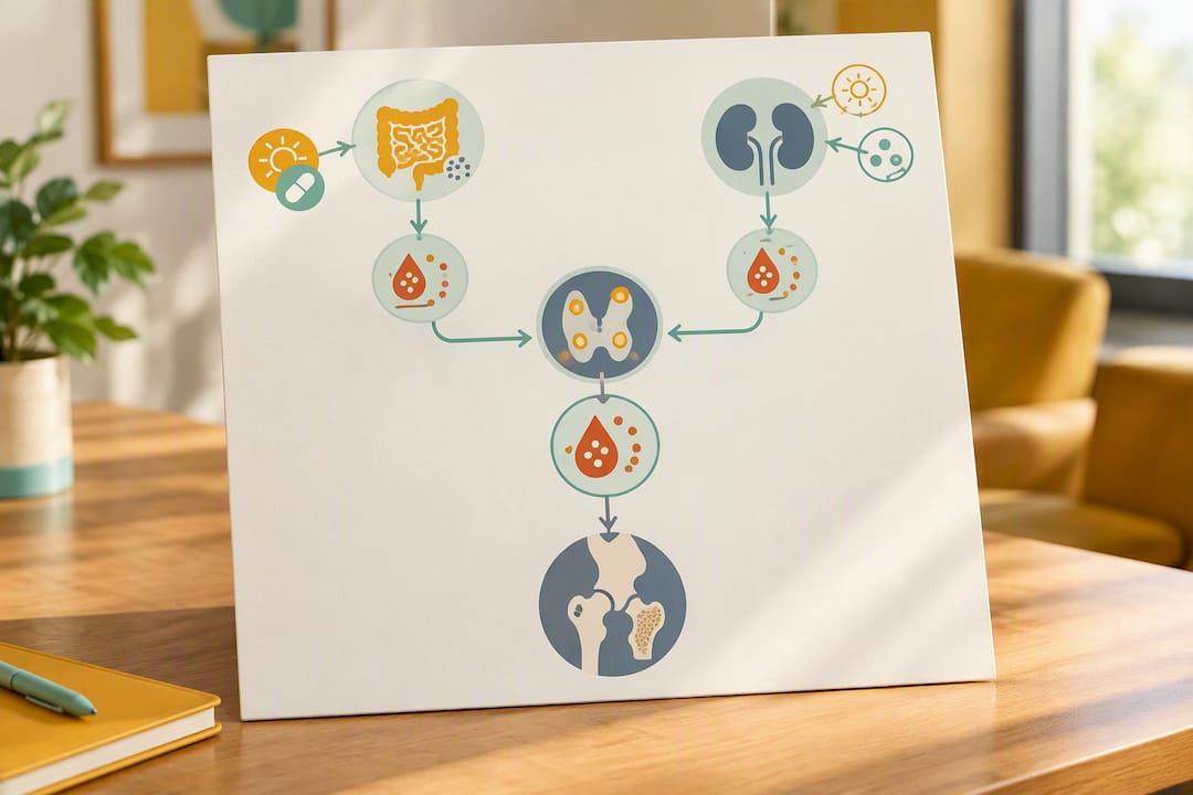
In vitamin D deficiency, low calcium absorption from the gut triggers the parathyroid glands to compensate by secreting more PTH, which pulls calcium from bone to keep blood levels stable. In CKD, failing kidneys can’t activate vitamin D properly and struggle to excrete phosphate, which together drive PTH levels up chronically, a process tied to what nephrologists call renal osteodystrophy. Both pathways lower hip bone density over time, though the mechanisms behind the damage differ from the direct, tumor-driven overproduction seen in primary disease.
Management differs accordingly. Vitamin D deficiency responds to repletion, correcting the deficiency directly addresses the compensatory PTH rise. CKD related secondary hyperparathyroidism is more complicated, since surgery is rarely the first move and the focus shifts to phosphate control, vitamin D analogs, and sometimes cinacalcet, all managed within a nephrology framework rather than an endocrine surgical one. Bone density monitoring remains important in both cases, but the treatment target is the underlying cause, not the parathyroid gland itself.
What’s Actually Happening at the Cellular Level
PTH’s relationship with bone is genuinely two-sided, and understanding that duality explains a lot about why hyperparathyroidism causes damage while certain PTH-based drugs are used to treat osteoporosis.
At the cellular level, PTH binds to receptors on osteoblasts, the bone-building cells, which then signal osteoclast precursors to mature and begin resorbing bone. Continuous, unrelenting PTH exposure, the kind seen in untreated primary hyperparathyroidism, pushes this system firmly toward resorption. Osteoclasts stay chronically activated, chew through cortical bone faster than it can be replaced, and the net effect over months and years is measurable bone loss concentrated at cortical sites.
Intermittent, low-dose PTH exposure does the opposite. This is the basis for teriparatide, an anabolic osteoporosis drug that delivers PTH in brief daily pulses rather than a constant stream. Given intermittently, PTH favors osteoblast activity and new bone formation over resorption. It’s the same hormone, the same receptor, producing opposite skeletal outcomes depending entirely on the pattern of exposure.

This dual nature is why untreated hyperparathyroidism and PTH-based osteoporosis treatment can look, on paper, like they involve the same hormone doing contradictory things. They’re not contradictory at all. Context and timing determine whether PTH builds bone or breaks it down, and that distinction is central to why continuous overproduction from a parathyroid tumor causes the specific, cortical-dominant damage pattern seen in this disease.
Fracture Risk Tools: Why Standard Calculators Can Miss the Danger
Standard fracture risk calculators, including the widely used FRAX tool, were built primarily on data from postmenopausal osteoporosis, not primary hyperparathyroidism. That mismatch creates a real blind spot for clinicians trying to gauge risk in this specific patient population.
FRAX estimates 10-year fracture probability using factors like age, prior fracture history, and femoral neck BMD, but it doesn’t account for the cortical-dominant damage pattern or the DXA discordance that defines hyperparathyroidism. A patient can generate a low or moderate FRAX score using spine or femoral neck BMD while actual cortical bone quality at the wrist tells a much more concerning story.
This is exactly why TBS and multi-site DXA carry extra weight in this specific condition. Clinicians increasingly treat hyperparathyroidism as a case where standard risk calculators need supplementing rather than replacing, layering TBS results and radial bone density onto the conventional FRAX output to get a fuller risk picture. A patient with a reassuring FRAX score but a low TBS or a low radial T-score is not necessarily a low-risk patient, and treating them as one based on the calculator alone can miss real, actionable fracture risk.
For patients, the practical takeaway is straightforward: a good FRAX score in the context of confirmed hyperparathyroidism shouldn’t be taken as the final word. It’s one data point among several, and in this disease, it’s arguably not the most reliable one.
Ruling Out Look-Alike Conditions
Elevated PTH with bone loss can resemble several other metabolic bone disorders, and getting the diagnosis right changes the entire treatment path.
Osteomalacia, caused by severe vitamin D deficiency, produces soft, poorly mineralized bone and can elevate PTH secondarily, but it responds to vitamin D repletion rather than parathyroid surgery. Paget’s disease of bone creates localized areas of abnormal, disorganized remodeling that can look like focal bone loss on imaging but follows a completely different pattern and treatment protocol, typically bisphosphonates targeted at the affected site. Multiple myeloma, a blood cancer, can cause both elevated calcium and bone lesions that mimic hyperparathyroidism’s biochemistry, which is why unexplained hypercalcemia with bone pain often triggers additional lab work to rule out malignancy before settling on a parathyroid diagnosis.
The distinguishing lab pattern for primary hyperparathyroidism is elevated or inappropriately normal PTH paired with elevated calcium. Osteomalacia typically shows low or normal calcium with low vitamin D. Paget’s disease usually presents with normal calcium and PTH but a markedly elevated alkaline phosphatase. Getting this differentiation right matters because the treatments diverge sharply. Surgery helps a parathyroid tumor. It does nothing for osteomalacia, and it’s the wrong answer entirely for myeloma.
What Happens If Hyperparathyroidism Goes Untreated
Left untreated, primary hyperparathyroidism doesn’t stabilize. The parathyroid tumor keeps producing excess hormone, and the pattern of cortical bone loss described earlier continues accumulating year after year, often without any obvious symptoms to signal that damage is progressing.
Long-term untreated disease is associated with a cumulative rise in fracture risk at the hip, spine, and wrist, driven by the same continuous resorption process, just given more time to do its damage. Bone that has thinned progressively over a decade doesn’t just stay thin; it becomes measurably weaker and more porous, and each additional year of unchecked PTH exposure adds to that structural deficit.
The condition also carries effects beyond the skeleton that compound the picture, including kidney stones from chronically elevated calcium, and a range of nonspecific symptoms, fatigue, mood changes, and cognitive fog, that patients sometimes attribute to unrelated causes for years before hyperparathyroidism gets diagnosed. Left unaddressed, elevated calcium can also affect cardiovascular health over the long term.
This is the strongest argument for early diagnosis and treatment: bone loss in this disease is progressive, not static. It doesn’t level off on its own the way some benign conditions do. Every additional year of untreated PTH excess is another year of cortical bone that has to be rebuilt later, and the surgical outcome data above suggests that rebuilding, while real and measurable, takes time the body doesn’t get back any faster for having lost it slowly.
A Checklist Worth Bringing to Your Next Appointment
The core answer here doesn’t change much no matter how deep you go into the mechanisms: excess PTH damages cortical bone in a fairly predictable pattern, and for most surgical candidates, that damage starts reversing within the first year and keeps improving through the second. That’s genuinely good news, but it only helps if you’re asking your care team the right questions.
A few worth writing down before your next visit: Which DXA sites should be tested, and does that include the 1/3 radius? When will follow-up scans happen, and is 6 months too early to expect a meaningful change? Given my baseline PTH and calcium levels, how much BMD recovery is realistic for someone like me? Should antiresorptive therapy start now, or is it better to wait and see what surgery accomplishes on its own?
Bone recovery after hyperparathyroidism treatment is a marathon tracked in months, not a sprint measured in weeks, and the patients who navigate it best tend to be the ones asking specific, site-by-site questions rather than accepting a single vague number. Integrated care, endocrinology, sometimes nephrology, and supportive services working from the same monitoring plan, tends to produce clearer answers than any one piece of the puzzle alone.
— Aaron
Supporting Your Bones Between Medical Visits
Between DXA scans and endocrinology appointments, there’s a real gap where patients often wonder what else they can do. Supervised, once-weekly osteogenic loading sessions apply controlled mechanical force intended to stimulate a bone-building response, paired with supportive modalities like red light therapy, PEMF mats, BioCharger sessions, vibration plates, and hydromassage lounges for recovery and overall wellness.

These sessions are designed as an adjunct alongside the medical or surgical care your endocrinologist or surgeon has already prescribed, not a replacement for it. If you have active hyperparathyroidism, talk to your treating physician before starting any new loading program, especially while calcium levels are being managed. For patients cleared to add a mechanical bone-loading component to their routine, Osteostrong’s centers offer a way to explore what a supervised, once-weekly session actually involves. Check with your local center about scheduling an initial assessment to see whether it fits into your current treatment plan.
Sources
- Changes in bone mineral density after surgical intervention …
- Primary hyperparathyroidism: pathophysiology and impact on bone
- Trabecular bone score and its utility in fracture risk assessment
- Factors influencing bone mineral density in hyperparathyroidism phenotypes: a prospective study
FAQ
Should you take vitamin D if you have hyperparathyroidism?
Vitamin D repletion is often necessary, but only under a clinician’s guidance, since correcting a deficiency in someone with primary hyperparathyroidism can occasionally raise calcium further. Your endocrinologist should set the target level and monitor calcium closely while you adjust your dose.
What are early warning signs of parathyroid problems?
Early signs often include fatigue, bone or joint aches, kidney stones, excessive thirst, and mood changes like irritability or brain fog. Many people have no symptoms at all and are diagnosed only after a routine blood test shows elevated calcium.
What does hyperparathyroidism do to your teeth?
Chronic PTH excess can cause loss of the lamina dura, the thin bone layer around tooth roots visible on dental X-rays, and can contribute to loose teeth or jawbone changes in advanced, longstanding cases. Dentists sometimes spot this pattern before a medical diagnosis is made.
What can trigger hyperparathyroidism?
Primary hyperparathyroidism is usually caused by a benign parathyroid adenoma that develops for unclear reasons, though prior neck radiation and certain rare genetic syndromes raise the risk. Secondary hyperparathyroidism is different. It’s typically triggered by vitamin D deficiency or chronic kidney disease driving compensatory PTH increases.
How much bone density improvement is realistic after parathyroidectomy?
Many patients see spine and hip BMD improvements starting around 6 months after surgery, with continued gains through 24 months. One prospective cohort found about 40% of patients gained more than 5% BMD at the spine, though individual results vary based on baseline PTH severity and other health factors.