

Menopause triggers a fast, measurable drop in bone density that raises your fracture risk for the rest of your life, and the earlier you act, the more skeleton you keep. That’s the entire premise of this guide, so here’s what to do this month, not “someday”:
- Pull up your family and personal risk factors (early menopause, low body weight, prior fractures, smoking).
- Ask your doctor whether you qualify for a baseline DXA scan now rather than waiting until 65.
- Start weight-bearing or resistance training this week; bone responds to mechanical stress, not intention.
- Check whether your calcium and vitamin D intake actually hits the targets researchers use, not just what “feels like enough.”
Groups like SWAN and the NIH have tracked this transition in thousands of women, and the DXA scan remains the tool that turns “probably at risk” into an actual number.
Key Takeaways
Menopause accelerates bone loss through an estrogen-driven imbalance between bone breakdown and bone building, and combining early screening, targeted loading, and guideline-based treatment is the most reliable way to protect against fracture.
| Point | Details |
|---|---|
| Loss is front-loaded | Most rapid bone loss happens in the year before and two to three years after your final period. |
| Get a baseline DXA | Ask about screening earlier than 65 if you have risk factors like early menopause or a fragility fracture. |
| Load the skeleton directly | Progressive resistance training and weight-bearing activity provide a stronger bone stimulus than cardio alone. |
| Monitor even on treatment | Up to 15% of women keep losing bone on hormone therapy, so repeat DXA scans matter regardless of treatment. |
| Add targeted mechanical loading | Osteostrong’s supervised osteogenic loading sessions complement medical care with objective progress tracking. |
Table of Contents
- What Causes Menopause Bone Loss?
- Who Loses Bone the Fastest After Menopause?
- How Do Doctors Diagnose Bone Loss and Fracture Risk?
- What Actually Prevents Bone Loss During Menopause?
- When Should You Consider Medication for Bone Loss?
- Can You Reverse Bone Loss After Menopause?
- How Often Should You Recheck Your Bone Density?
- Does Mechanical Loading Really Help Build Bone?
- A Publisher’s Take on Lifelong Bone Strategy
- Ready to Add Targeted Loading to Your Bone Health Plan?
- Frequently Asked Questions
- Sources
What Causes Menopause Bone Loss?
Estrogen keeps a constant construction project in balance. Cells called osteoclasts dissolve old bone, cells called osteoblasts rebuild it, and estrogen normally keeps the demolition crew from outpacing the builders. When estrogen drops during the menopause transition, osteoclasts get more active and live longer, while osteoblast output doesn’t scale up to match. The result is a net loss of bone tissue, month after month, concentrated in the years surrounding your final period.
This isn’t a slow, even decline that started in your 30s and just continues. It’s a distinct acceleration. Research on primary osteoporosis in postmenopausal women shows women can lose roughly 10% to 20% of their bone density in the 5 to 7 years around menopause, a window most women spend focused on hot flashes and sleep, not their skeleton.
- The fastest phase starts about a year before your final menstrual period and runs another two to three years past it.
- During that window, annual losses average around 2%, according to SWAN’s bone health data, with some cohorts showing a notable cumulative decline over a decade.
- Trabecular bone (the spongy tissue inside the spine and hip) loses density faster than cortical bone (the dense outer shell of long bones), which is part of why spine fractures often show up before hip fractures in early postmenopausal years.
Bone loss snapshot: In the fast-loss window alone, women can lose about 2% of bone density per year, with the spine typically losing ground faster than the hip, per SWAN.
The clinical stakes here aren’t abstract. Population data on osteoporosis pathophysiology links these bone mineral density losses directly to higher fracture rates, and bone lost during this narrow window is harder to rebuild later. That’s why intervention timed to the transition, not years after it, protects the internal scaffolding (bone microarchitecture) that keeps bone strong even after some density is gone.
Who Loses Bone the Fastest After Menopause?
Some women sail through menopause with modest bone loss. Others lose density fast enough to develop osteoporosis within a few years. The difference usually comes down to a specific set of risk factors clinicians watch for:
- Early menopause (before age 45) or surgical removal of the ovaries, which extends your total years of estrogen deficiency.
- Low body weight or a small frame, since there’s less bone reserve to draw from.
- A family history of hip fracture, particularly in a parent.
- Smoking and heavy alcohol use, both of which interfere with bone remodeling.
- Long-term glucocorticoid (steroid) use, malabsorption conditions like celiac disease, and thyroid or parathyroid disorders.
Risk also varies by ethnicity. Women of Asian and White ancestry tend to show higher rates of osteoporosis diagnosis, while Black women generally have higher baseline bone density but still face serious fracture consequences when bone loss does occur. None of this is destiny. Body weight, smoking, and alcohol use are modifiable; age at menopause and family history are not.
A fragility fracture (breaking a bone from a fall at standing height or less), noticeable height loss, or a new stooped posture (kyphosis) are red flags that warrant evaluation now, not at your next annual physical.
How Do Doctors Diagnose Bone Loss and Fracture Risk?
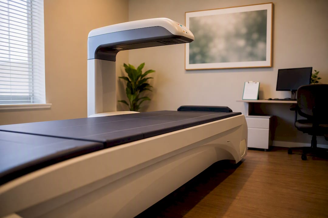
A DXA scan (dual-energy X-ray absorptiometry) is the standard for diagnosing bone loss, and most guidelines recommend one for all women by age 65, earlier if you carry risk factors like early menopause or a fragility fracture. The scan produces a T-score, which compares your bone density to that of a healthy young adult.
| T-score | Category | Clinical implication |
|---|---|---|
| Normal | No treatment typically needed; recheck per risk profile | |
| Osteopenia (low bone mass) | Monitor; prevention and lifestyle changes emphasized | |
| Osteoporosis | Fracture risk significantly elevated; treatment often indicated |
The T-score alone doesn’t tell the whole story. That’s where FRAX (the Fracture Risk Assessment Tool) comes in. FRAX combines your bone density with clinical factors like age, weight, smoking status, and fracture history to estimate your 10-year probability of a hip or major osteoporotic fracture. Clinicians use this combined picture, not the T-score in isolation, to decide whether medication makes sense.
A typical screening path looks like this:
- Baseline DXA measuring both the lumbar spine and femoral neck, since results between sites can diverge meaningfully.
- Blood work to rule out secondary causes: calcium, vitamin D, thyroid-stimulating hormone (TSH), and celiac screening if malabsorption is suspected.
- A vertebral fracture assessment or spine X-ray if you’ve had unexplained height loss or back pain, since silent spine fractures are common and easy to miss.
- A FRAX calculation once DXA results are in hand, to translate the numbers into an actual treatment conversation.
Ask specifically about your spine and hip scores separately. Osteopenia at the hip with normal spine density (or vice versa) changes the urgency of follow-up.
What Actually Prevents Bone Loss During Menopause?
Prevention works on three fronts at once: feeding the bone-building process, loading the skeleton hard enough to trigger it, and reducing your odds of a fall in the first place. Skip any one of the three and you’re leaving protection on the table.
Nutrition targets. Women over 50 generally need more calcium than their younger selves, and Mayo Clinic’s bone health guidance points to adequate calcium plus sufficient protein as foundational, not optional. AAOS frames typical calcium needs at roughly 1,000 to 1,200 mg daily depending on age, with vitamin D supplementation recommended for anyone with confirmed insufficiency. Protein matters more than most women realize, since bone is partly protein by weight, and magnesium, vitamin K, and phosphorus all play supporting roles in mineralization that a calcium-only focus misses.
Exercise that actually loads bone. Not all movement counts equally here. Research on progressive resistance training shows high-intensity resistance work can increase bone density in postmenopausal women and may protect against low-trauma fractures, while also improving the balance that prevents falls in the first place. A useful weekly framework:
- Two to three sessions of resistance training targeting the hips, spine, and wrists, the sites most vulnerable to fracture.
- Weight-bearing impact activity (brisk walking, stair climbing, dancing) most days of the week.
- Balance and stability work, since a fall you avoid is a fracture you never have to treat.
Pro Tip: Swimming and cycling are excellent for your heart, but they’re largely non-weight-bearing, so they don’t deliver much of a bone-building signal. If bone density is your goal, pair cardio with resistance work rather than relying on it alone.
Round it out with the basics that rarely get airtime: quitting smoking, keeping alcohol moderate, and clearing home fall hazards like loose rugs, poor lighting, and unstable footwear.
When Should You Consider Medication for Bone Loss?
Medication enters the picture when fracture risk is high or very high, not automatically at the first sign of osteopenia. Clinical guidance generally classifies risk using fracture history, DXA results, and FRAX-based fracture probability to decide who benefits from drug therapy versus lifestyle measures alone.
Here’s how the major options compare on the dimensions that matter most:
- Menopausal hormone therapy (estrogen, with progestogen if you have a uterus) works best when started near menopause for women with vasomotor symptoms and no contraindications; it slows bone loss but around 5% to 15% of women may keep losing bone anyway, according to Cleveland Clinic, so monitoring still matters.
- Bisphosphonates (oral or IV) are typically first-line for osteoporosis, taken weekly, monthly, or annually depending on the drug; they reduce spine and hip fracture risk over one to three years and carry rare risks of atypical femur fracture and osteonecrosis of the jaw with long-term use.
- Denosumab (Prolia), an injection given every six months, works well for women who can’t tolerate bisphosphonates or have kidney impairment, though bone loss can rebound quickly if doses are stopped without follow-up therapy.
- Teriparatide (Forteo), a daily injection, is an anabolic agent (it builds new bone rather than just slowing loss) generally reserved for very high-risk women or those with prior fractures, typically used for no more than two years.
- Romosozumab (Evenity), a monthly injection, builds bone quickly for one year and is often followed by a bisphosphonate to preserve gains; it carries a cardiovascular warning that rules it out for women with recent heart attack or stroke.
Some women continue losing bone despite being on hormone therapy. That single fact is the strongest argument for objective DXA monitoring rather than assuming any treatment is working just because you’re taking it.
Before starting anything, your clinician should check kidney function, discuss how long you’ll likely need treatment, and talk through whether a “drug holiday” makes sense down the line, particularly for bisphosphonates.
Can You Reverse Bone Loss After Menopause?
Getting back to your peak, premenopausal bone mass generally isn’t realistic once significant loss has occurred. What’s genuinely achievable: stopping further decline and gaining back a meaningful margin of density, which is often enough to substantially cut your fracture risk.
- Menopausal hormone therapy mainly slows loss rather than rebuilding significant bone.
- Bisphosphonates and denosumab stabilize density and modestly increase it over time.
- Anabolic agents like teriparatide and romosozumab produce the largest measurable BMD gains.
- Supervised osteogenic loading and progressive resistance training build bone through mechanical stimulus, working alongside any medication rather than replacing it.
The real goal isn’t chasing a perfect T-score. It’s reducing your odds of the fracture that changes your independence.
How Often Should You Recheck Your Bone Density?
Most stable women repeat DXA scans every one to two years, though your clinician may space that out further once density holds steady on treatment. Anyone starting a new bone medication typically gets a follow-up scan around the one to two-year mark to confirm the drug is actually working.
- Repeat labs (calcium, vitamin D) periodically if you’re being treated for a deficiency.
- Get a vertebral imaging or spine X-ray sooner if you develop new back pain or notice height loss.
- Push for earlier reassessment if you have a new fracture, a surprising drop in DXA results, or side effects that make you want to stop treatment. None of those should wait for your next scheduled appointment.
Does Mechanical Loading Really Help Build Bone?
Bone responds to mechanical stress the way muscle responds to resistance training. High-intensity, site-specific loading is one of the most consistently supported stimuli for maintaining and improving density, according to AAOS, and it works alongside medication rather than against it.
The evidence has real limits. Trial sizes are often modest, and outcomes vary by how much force the loading actually delivers to the hip and spine, the two sites where fracture risk matters most.
- Supervised programs typically involve one weekly session with progressive intensity increases over time.
- Objective tracking (repeat DXA) confirms whether the loading is producing real density change, not just muscle strength.
- These sessions are designed to complement medical therapy and lifestyle work, not replace a doctor’s evaluation.
Given that even hormone therapy leaves 5% to 15% of women still losing bone, objective tracking through DXA remains the only way to know if any approach, mechanical or pharmaceutical, is actually working for you.
A Publisher’s Take on Lifelong Bone Strategy
We built Osteostrong’s approach around a simple observation: bone needs a strong, specific mechanical signal, and most women never get one after menopause hits. Weekly supervised osteogenic loading sessions are designed to deliver that stimulus safely, alongside modalities like PEMF and red light therapy that support recovery. None of this replaces your doctor. It’s a conversation worth having alongside one.
Ready to Add Targeted Loading to Your Bone Health Plan?
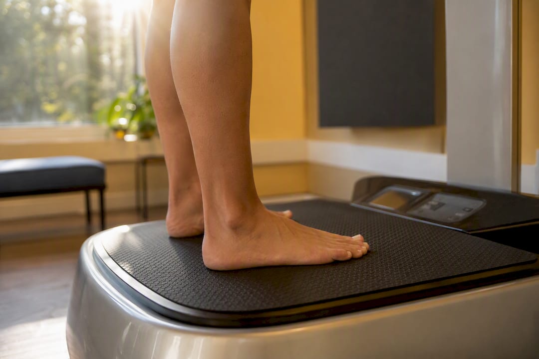
Nutrition and medication address two-thirds of the bone loss equation. The third piece, mechanical loading strong enough to actually signal new bone growth, is the part most home exercise routines never quite reach. Osteostrong’s weekly sessions use targeted, high-intensity loading equipment designed to deliver that stimulus in minutes, without the impact risk of high-intensity jumping programs.

If you’re already tracking your DXA results and wondering what more you can do between doctor visits, a session at a location like Osteostrong Rancho Cordova or Osteostrong Lake Mary takes under 15 minutes and comes with progress tracking built in. Book an introductory assessment to see how your current bone strength stacks up, and bring your last DXA report if you have one.
Frequently Asked Questions
How much bone loss is normal during menopause?
Losing roughly 10% to 20% of bone density over the 5 to 7 years surrounding menopause falls within the range seen in population studies, though the exact amount varies by individual risk factors and site (spine versus hip).
Can you reverse bone loss after menopause?
Full reversal to premenopausal bone mass isn’t typically realistic, but stopping further loss and gaining back a modest amount of density through treatment and targeted loading measurably reduces fracture risk.
How do you build bone density after menopause?
Combine adequate calcium and protein intake, vitamin D if you’re deficient, progressive resistance training, and, when fracture risk is high, a bone-specific medication chosen with your doctor based on your DXA and FRAX results.
Does menopause cause joint pain as well as bone loss?
Yes, declining estrogen affects joint tissue and inflammation levels, which is why many women notice new joint stiffness or aches alongside bone density changes during the transition, even though the two involve different tissues.
This article is general information, not a substitute for advice from a qualified doctor. Consult a qualified healthcare professional about your own circumstances before acting on anything here.
Sources
- Primary osteoporosis in postmenopausal women – PMC – NIH
- SWAN Fact Sheet: Bone Health over the Menopause Transition
- Osteoporosis: Pathophysiology, clinical consequences, and epidemiology – NCBI Bookshelf
- What To Know About Menopause and Bone Loss – Cleveland Clinic
Trackbacks/Pingbacks